Shetlands

— Aucanada —
Health is our the highest priority. It´s the reason why we test all our dogs of the most common genetic diseases within this breed. For thoses diseases that cannot be tested, we do a deep long study of the breeding lines we want to use, their combinations and individual health results.

Certified hip, elbow, shoulder x-rays (OCD) and spine (Spondylosis).

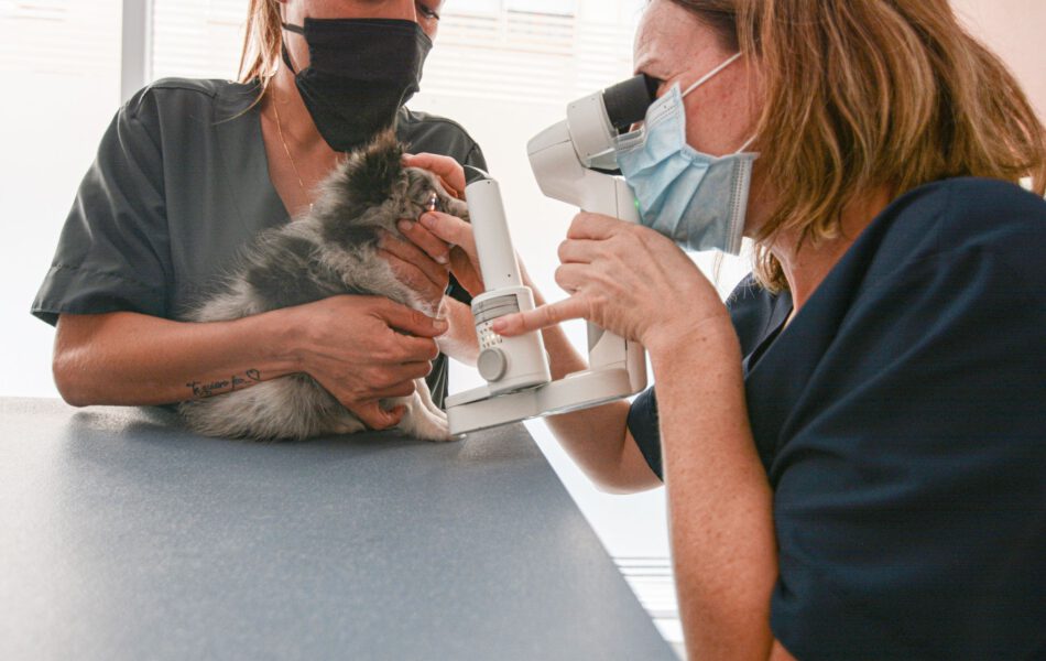
Eye exams by ECVO.

Genetically tested for:

CEA (Collie Eye Anomaly)

Sensitivity to ivermectin (defective MDR1 gene)

Canine degenerative myelopathy (DM)

Von Willebrand disease type 3 (vWDIII)

100%

Certified x-rays

Certified hip, elbow, shoulder x-rays (OCD).

100%

Eye exams

Eye exams by ECVO.

100%

Genetically tested

For CEA, MDR1, DM & vWDIII.

SHETLAND SHEEPDOG STANDARD
ORIGIN

Great Britain.

DATE OF PUBLICATION OF THE OFFICIAL VALID STANDARD

19.08.2013

UTILIZATION

Companion dog and Sheepdog.

CLASSIFICATION F.C.I.

Group 1 Sheepdogs and Cattle Dogs.

Section 1 Sheepdogs

Without working trial.

GENERAL APPEARANCE

Small, long-haired working dog of great beauty, free from cloddiness and coarseness, action lithe and graceful. Outline symmetrical so that no part appears out of proportion to whole. Abundant coat, mane and frill, shapeliness of head and sweetness of expression combine to present the ideal.

IMPORTANT PROPORTIONS

Skull and muzzle of equal length, dividing point inner corner of eye. Slightly longer from point of shoulder to bottom of croup than height at withers.

BEHAVIOUR/TEMPERAMENT

Alert, gentle, intelligent, strong and active. Affectionate and responsive to his owner, reserved towards strangers, never nervous.

HEAD

Head refined and elegant with no exaggerations; when viewed from top or side a long, blunt wedge, tapering from ear to nose. Width and depth of skull in proportion to length of skull and muzzle. Whole to be considered in connection with size of dog.

CRANIAL REGION

Skull: Flat, moderately wide between ears, with no prominence of occipital bone. Topline of skull parallel to topline of muzzle.
Stop: Slight but definite.

FACIAL REGION

The characteristic expression is obtained by the perfect balance and combination of skull and foreface, shape, colour and placement of eyes, correct position and carriage of ears.
Nose: Black.
Lips: Tight with black rims.
Jaws/Teeth: Jaws level, clean, strong with well-developed underjaw.
Teeth sound with a perfect, regular and complete scissor bite, i.e. upper teeth closely overlapping lower teeth and set square to the jaws. A full complement of 42 properly placed teeth highly desired.
Cheeks: Flat, merging smoothly into well rounded muzzle.
Eyes: Medium size obliquely set, almond-shape with black rims. Dark brown except in the case of merles, where one or both may be blue or blue flecked.
Ears: Small, moderately wide at base, placed fairly close together on top of skull. In repose, thrown back; when alert brought forward and carried semi-erect with tips falling forward.

NECK

Muscular, well arched, of sufficient length to carry head proudly.

BODY

Back: Level, with graceful sweep over loins.
Croup: Sloping gradually to rear.
Chest: Deep, reaching to point of elbow. Ribs well sprung, tapering at lower half to allow free play of forelegs and shoulders.

TAIL

Set low; tapering bone reaches to at least hock; with abundant hair and slight upward sweep. May be slightly raised when moving but never over level of back. Never kinked.

LIMBS

FOREQUARTERS:
General appearance : Forelegs straight when viewed from front, muscular and clean with strong, but not heavy, bone.
Shoulder: very well laid back. At withers, separated only by vertebrae, but blades sloping outwards to accommodate desired spring of ribs. Shoulder joint well angled.
Upper arm: Approximately equal in length with shoulder blade.
Elbow: Equidistant from ground and withers.
Metacarpus (Pastern): Strong and flexible.

HINDQUARTERS:
Thigh: Broad and muscular, thigh bones set into pelvis at right angles.
Stifle: Joint has distinct angle.
Hock joint: Joint clean cut, angular, well let down with strong bone. Hocks straight when viewed from behind.

FEET:
Oval, soles well padded, toes arched and close together.

GAIT/MOVEMENT

Lithe, smooth and graceful with drive from hindquarters, covering the maximum amount of ground with the minimum of effort. Pacing, plaiting, rolling, or stiff, stilted, up and down movement highly undesirable.

COAT

Hair:
Double; outer coat of long hair, harsh-textured and straight. Undercoat soft, short and close. Mane and frill very abundant, forelegs well feathered. Hindlegs above hocks profusely covered with hair, below hocks fairly smooth. Face smooth. The coat should fit the body and not dominate or detract from the outline of the dog. Smooth-coated specimens highly undesirable.

Colour:
Sable: clear or shaded, any colour from pale gold to deep mahogany, in its shade, rich in tone. Wolf-sable and grey undesirable.
Tricolour: intense black on body, rich tan markings preferred.
Blue Merle: clear silvery blue, splashed and marbled with black. Rich tan marking preferred but absence not penalised. Heavy black markings, slate or rusty tinge in either top or undercoat highly undesirable; general effect must be blue.
Black and White, and Black and Tan: also recognised colours. White markings may appear (except on black and tan) in blaze, collar and chest, frill, legs and tip of tail. All or some white markings are preferred (except on black and tan) but absence of these markings
not to be penalised. Patches of white on body highly undesirable.

SIZE

Ideal height at withers:
Males 37 cm
Females 35,5 cm
More than 2 1/2 cm above or below these heights highly undesirable.

FAULTS

Any departure from the foregoing points should be considered a fault and the seriousness with which the fault should be regarded should be in exact proportion to its degree and its effect upon the health and welfare of the dog.

DISQUALIFYING FAULTS

Aggressive or overly shy.
Any dog clearly showing physical or behavioural abnormalities shall be disqualified.

N.B.

Male animals should have two apparently normal testicles fully descended into the scrotum.
Only functionally and clinically healthy dogs, with breed typical conformation, should be used for breeding.

OFFICIAL CERTIFICATION

CEA (Collie Eye Anomaly) EVG – UCM – LABOKIN
Sensitivity to ivermectin (defective gene MDR1) EVG – SLOVGEN
Von Willebrand disease type 3 (vWDIII) EVG – SLOVGEN
Canine degenerative myelopathy (DM) EVG – UCM – OFA

Entities where the radiographs are certified:
Hips and Elbows: AVEPA – OFA – SETOV
Patella Luxation & shoulders(OCD): OFA
Spondylosis: SETOV
Official  Organization  ophthalmological certificate: ECVO

HEALTH
Genetics
CEA (Collie eye anomaly)

Collie eye anomaly – characteristics

Collie eye anomaly (CEA) is a complex hereditary ocular disorder which is characterized by regional hypoplasia of choroids, the highly vascularized layer of the eye that supplies blood and nutrients to the retina. Occasionally, tortuous retinal vessels and multiple retinal folds are also observed. The clinical symptoms can vary greatly among affected dogs within one breed, between parent and offspring and within a litter. No medical treatment of the disease is available.

CEA is widespread in dogs, with a prevalence of 65–97% for rough and smooth collies, while the prevalence in Border collies is lower. CEA is also recognized in all herding dogs or breeds that are derived from classic herding breeds.

CEA genetics

CEA inheritance is autosomal recessive with variable expression and pleomorphism (wide range in the clinical expression of the defect). Some mildly to moderately CEA-affected dogs appear to retain their normal vision function throughout their life. Severely affected individuals, particularly those with colobomas, can develop retinal detachments leading to blindness. In these cases, subretinal and preretinal neovascularization and intraocular haemorrhage can occur.

MDR-1 (Multidrug resistance 1)

Multidrug resistance 1, also called MDR1, is an inherited condition affecting several breeds of dogs, especially herding dogs such as the Shetland sheepdog. The Mutation in the ABCB1 gene associated with MDR1 causes dysfunction of P-glycoprotein, which is responsible for removing certain drugs and toxins from the body. Clinical signs are most commonly associated with distribution of the drug in the central nervous system. MDR1 is inherited in an autosomal incomplete dominant manner in dogs meaning that dogs only need to inherit one copy of the mutated gene to be at an increased risk of developing adverse reactions to certain medications. Though adverse reactions to certain drugs are most commonly seen in dogs having two copies of the mutated gene, Carrier dogs can also experience drug sensitivities and dosages need to be adjusted accordingly. Thus, dogs that have one or two copies of the mutation are considered at-risk for adverse drug reactions. If an at-risk dog is treated with one of several common drugs (see below*), they are at risk of developing neurologic symptoms that could range from tremors, excess salivation, anorexia, and blindness to coma and even death. Because of the defective ability to metabolize specific drugs, these drugs can be lethal even at low doses. The MDR1 mutation does not cause adverse effects in dogs unless the dog is exposed to these drugs. Therefore, veterinarians should be notified when a dog is at risk for multidrug resistance 1 prior to administration of any medications.

DM (Degenerative Myelopathy )

Degenerative Myelopathy caused by Mutation of the SOD1 gene is an inherited neurologic disorder of dogs. This mutation is found in many breeds of dog, including the Shetland sheepdog. While it is not clear for some of the other breeds, Shetland sheepdogs are known to develop degenerative myelopathy associated with this mutation. The variable presentation between breeds suggests that there are environmental or other genetic factors responsible for modifying disease expression. The average age of onset for dogs with degenerative myelopathy is approximately nine years of age. The disease affects the White Matter tissue of the spinal cord and is considered the canine equivalent to amyotrophic lateral sclerosis (Lou Gehrig’s disease) found in humans. Affected dogs usually present in adulthood with gradual muscle Atrophy and loss of coordination typically beginning in the hind limbs due to degeneration of the nerves. The condition is not typically painful for the dog, but will progress until the dog is no longer able to walk. The gait of dogs affected with degenerative myelopathy can be difficult to distinguish from the gait of dogs with hip dysplasia, arthritis of other joints of the hind limbs, or intervertebral disc disease. Late in the progression of disease, dogs may lose fecal and urinary continence and the forelimbs may be affected. Affected dogs may fully lose the ability to walk 6 months to 2 years after the onset of symptoms. Affected small breed dogs, such as the Shetland sheepdog, often progress more slowly than affected large breed dogs and owners may postpone euthanasia until the dog is paraplegic.

PRA (Sheltie Progressive Retinal Atrophy )

Sheltie Progressive Retinal Atrophy is an inherited disorder that belongs in group of eye disorders known as progressive retinal atrophy. Progressive retinal atrophy (PRA) includes autosomal recessively inherited diseases that lead to degeneration of retinal photoreceptor cells in dogs and other pets. It is a canine equivalent to retinitis pigmentosa (RP), an inherited eye disorder affecting human beings. The first case of PRA was observed in England more than hundred years ago.

In general, PRA is characterized by disturbance of vision in dark, visual field defects, and abnormalities in the electroretinogram. It appears in both eyes simultaneously. The age of onset and rate of retinal degeneration varies between the different forms of the conditions. Some forms of PRA are common to multiple dog breeds, while others are recognized in just a single breed. Until now, it has been identified in more than 100 dog breeds. Almost all forms of PRA are inherited in a recessively manner, with exceptions in some breeds such as Old English Mastiffs, Bullmastiffs, Siberian Husky and Samoyed where forms of PRA are inherited as dominant or X-linked disorders.

There are two types of photoreceptors in the eye, rods and cones. Rods have an important role for vision in dim light and also night vision. PRA causes rode degeneration and in this way leads to night blindness. Researchers estimated that PRA causes death of around 95% of the dog’s photoreceptors. Dog owners or breeders recognize PRA by ”glow” or ”increased shine” in the eyes. The disease is progessive. Initial stage is characterized by night blindness, while advanced stage of PRA can cause full blindness in the dog. After the appearance of first symptoms, it usually takes one year for the disorder to progress from initial to the advanced stange, causing severe and irreverisble damage to the dog’s eye.

Sheltie Progressive Retinal Atrophy (CNGA1-PRA) is a form of PRA with an adult onset, with average of developing first symptoms at 5 years of age.

VWDIII (Von Willebrand disease type 3 )

Von Willebrand disease (vWD) is a bleeding disorder of variable severity that results from a quantitative or qualitative defect in von Willebrand factor (vWF). On vascular injury, vWf mediates platelet adhesion to exposed subendothelium and is involved in platelet-to-platelet aggregation. The disease is the most common hereditary bleeding disorder and it is genetically and clinically heterogeneous. Clinical signs of the disease include spontaneous bleeding from mucosal surfaces and excess blood loss after surgery or trauma. Three clinical types, 1, 2, and 3, have been described.

Von Willebrand disease type 3 is the severest form of the disease. It is inherited as an autosomal recessive trait, and affected individuals have no detectable vWF in their blood. Serious bleeding episodes require transfusions of blood or cryoprecipitate to supply the missing vWF. Heterozygous carriers have moderately reduced factor concentrations but generally appear to have normal hemostasis.

Testing of affected dogs can be done by von Willebrand disease type 3 antigen testing or by coagulation assays, but these procedures yield variable results. This variability makes it difficult for breeders to use this information to eliminate the disease-causing allele from their lines. Thus, it is highly desirable that DNA tests are run to get definitive answers.

Joints
HIPS DYSPLASIA

Hip dysplasia is a deformity of the hip that occurs during growth. The hip joint is a ball and socket joint. During growth, both the ball (the head of the femur, or thighbone) and the socket in the pelvis (acetabulum) must grow at equal rates.

In hip dysplasia, this uniform growth during puppyhood does not occur. The result is laxity (looseness) of the joint, followed by degenerative joint disease (DJD) or osteoarthritis (OA), which is the body’s attempt to stabilize the loose hip joint.

The degree of lameness that occurs is dependent on the extent of these arthritic changes and may not be correlated with the appearance of the hip joint on X-rays. Some pets with significant signs of hip dysplasia or osteoarthritis on X-rays may not exhibit any clinical signs, while others with minimal changes may experience severe pain and lameness.

Hip dysplasia is a genetic disease that is affected by factors such as diet, environment, exercise, growth rate, muscle mass, and hormones. As this disease is most commonly seen in large breed dogs (generally greater than 50 lbs or 22 kg), these puppies should be kept at a normal, lean weight during growth, rather than overfed and encouraged to grow “big.”


Hip dysplasia is a genetic disease that is affected by factors such as diet, environment, exercise, growth rate, muscle mass, and hormones.

ELBOWS DYSPLASIA

Elbow dysplasia is a painful condition that causes one or both elbows to develop abnormally while a puppy is growing. There are three main areas inside the elbow joint that can be affected; some dogs have just one problem area, while others suffer with a combination.


Elbow dysplasia causes pain, swelling, instability and often leads to arthritis.

Illustration to show elbow joint in dogs

The elbow is made of three bones: radius, ulna and humerus.


Symptoms of elbow dysplasia

Limping or stiffness (usually worse after exercise and difficult to spot if both elbows are affected)

Less enthusiasm to go for walks or play

Front paws pointing outwards and/or elbows held at a strange angle

Swollen, puffy elbows (in severe cases)

Most dogs start showing symptoms between 5 – 18 months old. However, dogs with mild elbow dysplasia may not show signs until later in life, once they have developed arthritis in their elbows.

OCD (Shoulders)

Osteochondrosis is a condition that affects growing cartilage. The condition primarily affects young dogs going through rapid growth periods. Bone growth begins with the formation of cartilage, which then ‘ossifies’ or converts to bone. In osteochondrosis, this conversion process is abnormal, which leads to increased cartilage thickness where the bones join. The irregularly thickened cartilage suffers from poor blood supply and becomes softer and more susceptible to the effects of weight bearing impact. This leads to tissue formation, which sometimes progresses to the formation of cartilage flaps. Joint fluid then reaches the deeper layers of cartilage and bone, which creates inflammation. This inflammation causes pain and can lead to arthritis.

PATELLA LUXATION

In the patellar luxation, the kneecap pops out of its place – the femoral trochlea, causing pain, swelling and functional weakness in the dog, with the medial being the most frequent. It occurs more in females than in males, especially in small breeds and toy breeds, including the Shetland Sheepdog. It is a pathology that is characterized by poor alignment of the limb, deformations occur during the development of the animal, which cause the patella to slip out of place. It may be due to a congenital disease or, in some cases, caused by a trauma. It is convenient that the dogs that suffer this pathology congenitally, are not used in reproduction as it’s known that heritability can be transmitted through the generations.

Grade 1: Luxation with manual pressure – usually not painful.  Grade 1 luxations usually do not require any specific treatment but do need to be monitored for worsening. Sometimes pain medication is required.

Grade 2: Patella luxation with flexion and extension of the knee but returns to the groove on its own occasional pain is present.

Grade 3: Luxation with flexion and extension and returns to the groove with manual reduction considerable pain and lameness (limping).

Grade 4: The patellas are permanently luxated – severe condition. Grade 4 luxations usually require reconstructive surgery by a surgical specialist.

SPONDYLOSIS

Spondylosis is a disease of the spine where bone spurs (osteophytes) develop around the edges of the vertebrae. In order to diagnose this condition the spinal structure must be examined through the use of an X-ray or MRI. Most often spondylosis is diagnosed incidentally when an X-ray is taken of a pet for an unrelated reason. While evaluating the entire radiograph, a veterinarian would be able to see abnormal bone growth surrounding the spine. A normal spine will have individual “blocks” of vertebrae with the space in-between composed of the intervertebral disc. With spondylosis, bone spurs on the ends of the vertebrae are visible and, in more severe cases, the new bone growth can form a bridge above and below fusing or linking the vertebrae.